Testis Histology Diagram
Testis histology mcgraw companies tubules seminiferous Histology testes epididymis testis tunica vasculosa albuginea leydig cells seminiferous tubule Histology of testis
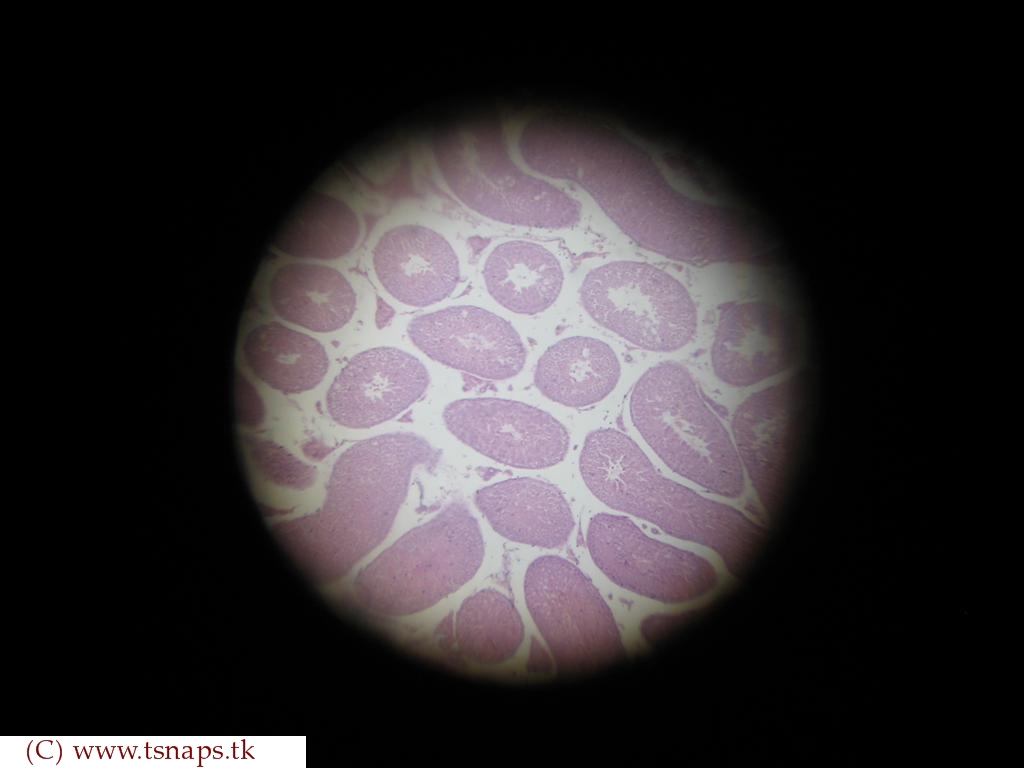
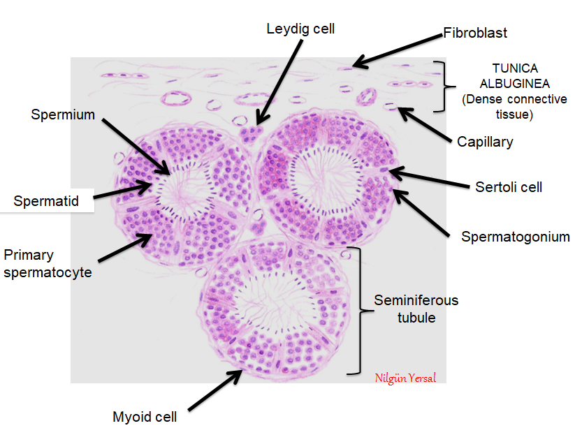
A cross section of the testis showing Sertoli cells and Leydig cells
Answer the following question. explain the histological structure of Histology testes reproductive male system ppt repro capsule fibrous tissue powerpoint presentation connective Testis epididymis normal histology pathweb nus annotations
Testis tubules seminiferous histology convoluted x10 male reproductive system file embryology young resolutions other preview size unsw med edu au
Testes testis diagram histological histology slidesTestis histology cgtrader turbosquid histologically Histology epididymis testesHistology testis slides testes.
Testis structure histology histological biology question explain answer following shaalaaTestis histology shaalaa biology Histology of testes & epididymisTestis, ductus deferens, and seminal vesicle histology.
Male reproductive system
Testis leydig cell seminiferous tubules tumor figureEpididymis testis histology nus pathweb annotations expand Histology testis diagram slides high resolutionHistology of testis by dr mohammad manzoor mashwani.
Testis histology labelled describe shaalaaLeydig cell tumor causes, symptoms, diagnosis, treatment & prognosis Gross anatomy and histology of testesTestes anatomy gross histology.

File:testis histology 004.jpg
Histology slides database: human testis histology slides (testes)Testis and epididymis – normal histology – nus pathweb :: nus pathweb Histology testis manzoorLeydig sertoli testis.
Testis histologyHistology slides database: histological diagram of testis (testes) Male reproductive: the histology guideHistology slides database: january 2014.

Histology of the testis
File:testis histology 1.jpgA cross section of the testis showing sertoli cells and leydig cells Testis and epididymis – normal histology – nus pathweb :: nus pathwebDescribe the t.s. of human testis.
Testis histologyTestis histology development spermatozoa embryology gametogenesis file human spermatogonia cells sertoli puberty pre postnatal sexual practical differentiation tubule seminiferous section Histology testis seminiferous anatomy male reproductive system tubules cell slides human file embryology medical lab epididymis testicular tissue frog tubuleHistology of testis.

Histology of testes & epididymis
Testis male histology reproductive labeled epididymis ducts system genital testes diagram labels gif accessory result leeds guide ac3d section testis anatomy histology Deferens ductus seminal testis vesicle histology do continue learningTestis male eosin hematoxylin reproductive system histology penis drawings vesicle seminal sperm.
File:testis histology 001.jpgTestis histology file embryology edu stain resolutions other preview size Describe the histology of testis with help of labelled diagramFile:testis histology 006.jpg.

Histology testis
.
.

Male Reproductive System

Histology of testes & epididymis

Male reproductive: The Histology Guide

Testis and Epididymis – Normal Histology – NUS Pathweb :: NUS Pathweb

A cross section of the testis showing Sertoli cells and Leydig cells

File:Testis histology 004.jpg - Embryology